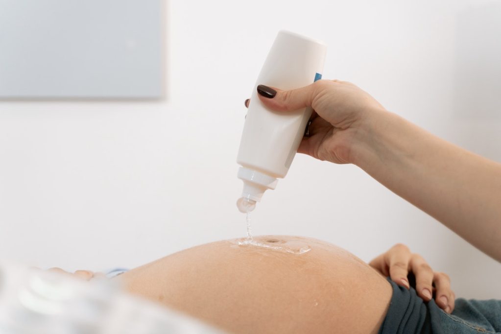
Sonograms can be a highlight of pregnancy. It’s a chance for an expecting parent or parents — and perhaps even other support people like grandparents — to catch a glimpse of the developing baby on the inside. It’s a reminder of why you’re enduring all those pregnancy symptoms.
During scans, you may note how incredible it is to see how fast the future baby is growing from a tiny little spec on a screen into a little one-to-be with a head, shoulders, knees, and toes.
Typically, these sonograms are two-dimensional. You can see some semblance of a developing human in black and white. It may feel underwhelming, especially if you log onto social media and see a friend post about a 3D sonogram. You may see the baby’s skin and more prominent facial features. Are 3D sonograms safe, and should you get one? Here’s what to know.

What is a 3D sonogram?
A 3D ultrasound uses high-frequency sound waves to show providers, patients, and partners images of a fetus’ body, including organs and tissues. That part is no different from the standard 2D sonogram. The distinction comes in the picture quality. A 3D ultrasound is sharper. You may see more features, and the images look more like the baby you’ll soon welcome.
Like a 2D sonogram, these scans are typically done abdominally. You can undergo one during any point of a pregnancy, though the baby generally is most ready for their close-up early in the third trimester, between 28 and 34 weeks. At that time, the fetus has developed enough that you may be able to pick up on whether your baby-to-be has high cheekbones or hair, but there’s still enough room in utero for them to move around and work their angles.

Benefits of a 3D sonogram
These sonograms, sometimes ordered for medical reasons, became popular among expectant parents during the pandemic. With OBGYN offices restricting support individuals within the office, some pregnant people booked a private 3D sonogram to allow partners or other family members to meet the developing baby.
With restrictions likely lifted at your provider’s office, should you still opt for a 3D sonogram? In some cases, your doctor may recommend it. For example, 3D ultrasounds are better at picking up issues like cleft palates that 2D technology simply cannot. The CDC notes that diabetes, smoking, and some medications increase the risk for cleft palates. Your provider will go over your risk factors and recommend a 3D scan if they believe it is necessary. Insurance should cover it, though it’s best to call to ensure that is the case.
Otherwise, the scans can be fun for people to see their baby-to-be more clearly, and the photos can be a keepsake for a nursery.

Are 3D sonograms safe?
The FDA says that sonograms are generally safe but discourages keepsake scans that aren’t medically indicated, citing unknown effects of ultrasound waves on a developing baby.
“Ultrasound energy has the potential to produce biological effects on the body. Ultrasound waves can heat the tissues slightly,” stated the FDA on imaging, which was last reviewed in September 2020, and reads, “In some cases, it can also produce small pockets of gas in body fluids or tissues (cavitation). The long-term consequences of these effects are still unknown.”
Insurance does not cover keepsake ultrasounds.

The bottom line on 3D sonograms
Sometimes, 3D sonograms may be medically indicated, such as to check for a cleft palate. In this case, your doctor will explain why and address any safety concerns so you can make an informed decision. Though non-medically necessary, 3D ultrasounds can make for beautiful keepsakes, but it’s best to discuss the risks and benefits of them with your doctor.


